- FREELY available online or in PDF format. Print books can be purchased here.
- All royalties from book sales go to Cancer Research UK. Want to read for free but still donate to Cancer Research UK? Click here.
-

Chapter 5: Brake Failure
In the summer of 1952, around the time that Renato Dulbecco was kick-starting the animal virus field in his Caltech basement lab, one of his close friends and compatriots was on her way towards a discovery that would be equally momentous. Rita Levi-Montalcini, like Dulbecco an ex-student of Giuseppe Levi, the extraordinarily charismatic Professor of Anatomy in Turin, was boarding a flight from Rome to Rio de Janeiro, where she was to spend a few months in the lab of another of Levi’s protégés, Hertha Meyer. She was not alone. In her pocket, safely stashed in a small cardboard box with an apple as an in-flight snack, were two white mice, who had already traveled to Italy with Rita from her lab at Washington University in St. Louis, Missouri. Unfortunately for the mice, they were not travelling as pets; both carried tumours, vital components of the work Rita hoped to do in Brazil.
Unlike her friend Renato, who had moved to the United States at the same time to learn molecular biology with another Levi alumnus, Salvador Luria, Rita had continued to work in Giuseppe Levi’s own field of experimental neuroembryology. Her move to St. Louis in 1946 had been at Levi’s prompting, to the lab of Viktor Hamburger, who had developed a system to study how the growth and migration of nerve fibres was directed by the nonneuronal tissue of embryos. The experimental model used in the Hamburger lab was the embryonic chick limb, a firm favourite of embryologists for both prosaic and scientific reasons: eggs were cheap, it was easy to watch what was going on in them by cutting a window in the shell, and the chick embryos were sufficiently large to make surgery such as grafting possible. Work in the lab was founded on the phenomenon that if the chick limb bud, the centre from which the limb grew, was completely destroyed, the nerves that would have grown into the limb also died. Conversely, if an extra limb bud was grafted onto an embryo, new nerve fibres would grow towards and enter the transplanted limb.
Things did not go well to begin with, and Rita, a frequent visitor to Dulbecco and Luria in Bloomington, Indiana, was tormented by doubts about whether she was doing the right thing. In her autobiography, In Praise of Imperfection, she relates that she had
… an unconfessed envy for geneticists and microbiologists, whose work I believed was far more interesting and promising than that of neuroembryologists.… I did not hide from … Luria my distressing doubts about the validity of our approach to the problem of the differentiation of the nervous system. I believe his doubts were much greater than mine, but he saw no way out of the difficulty. He excluded the possibility of my successfully taking up microbiology, unprepared as I was in the field, and encouraged me, without enthusiasm, to continue in my research (Levi-Montalcini 1988).
Rita’s frustration with her work lay in the sheer intractability of studying neuronal development; compared with the simplicity and solid theoretical grounding of molecular biology and genetics, it seemed a completely closed system. She could look at the growing nerves, by fixing and staining sections cut from the embryos, but there seemed no way of working out what was going on. However, in 1947, she made a conceptual breakthrough that completely restored her faith. Whilst examining sections cut from embryos at slightly different stages, she realised that she could track the temporal development of the nerve cells. As she says, “the startling realization that nerve cell populations were subject to quotas and to the elimination of excessive numbers in their ranks, as well as to migrations that went hand in hand with functional differentiation, showed that there were ontogenetic processes at work in the nervous system which were not as inaccessible to investigation as I had previously imagined” (Levi-Montalcini 1988).
In 1950, Hamburger received a letter from an ex-student of his, Elmer Bueker, which further galvanized Rita. Bueker was comparing the abilities of different mammalian tumours to grow as grafts on chick embryos, and had noticed that one tumour, S180, a mouse sarcoma, exerted an extraordinary effect on the nervous system of the host embryo. Nerve fibres originating from nearby sensory ganglia (clumps of nerve cell bodies) had grown into the tumour mass, completely innervating it just as if it were a grafted limb bud. Intrigued, Rita ordered in some S180-carrying mice and repeated Bueker’s experiment. What she saw was quite astonishing. “The mass of tumoral cells … were from all sides penetrated by bundles of brown-blackish nerve fibers. These fiber bundles passed between the cells like rivulets of water flowing steadily over a bed of stones.… I had the feeling that I had come upon a phenomenon without precedent in the rich case history of experimental embryology” (Levi-Montalcini 1988).
Rita repeated the experiment with a different mouse sarcoma, S37, and got the same results. Crucially, she then showed that even when she physically separated the tumours from the growing chick by grafting them outside the membrane encapsulating the embryo, the tumours still had the same effect. Rita realised to her great excitement that the tumour cells had to be releasing a soluble factor that was neurotropic, able to lure nerve fibres towards it, and that could also direct the future development of immature nerve cells, instructing them to differentiate into specific neuronal cell types. This factor, christened Nerve Growth Factor, soon abbreviated to NGF, turned out to be the key to the puzzle of how nonneuronal tissue could dictate how the nervous system developed.
Grafting tumours onto chick embryos was a cumbersome and slow technique, leading to Rita’s decision to go to Rio with her two mice carrying the S180 and S37 tumour cells. She had been hoping for an excuse to visit her friend Hertha Meyer in Brazil for some time, and this seemed perfect. Hertha had set up neuronal tissue culture in her lab, and growing the tumours with neurons in vitro was a far cleaner way to study the mystery factor than continuing with chicken eggs. There were some initial setbacks, but by the time of her return to St. Louis in January 1953, Rita had established tissue culture conditions under which nerve cells would put out dense, halo-shaped outgrowths of fibres in the direction of tumour cells. In a salutary lesson to today’s work-obsessed scientists, she also found the time to take a month’s holiday, starting first with Carnival in Rio and then going on to Peru and Ecuador.
Although Rita had set up a good assay for NGF in Rio, the obvious next step, attempting to purify the factor, was quite daunting, requiring a set of biochemical skills that neither she nor Viktor Hamburger possessed. However, in Rita’s absence, Viktor had made an inspired hiring decision—a young New Yorker called Stanley Cohen.
Stan Cohen had started his biochemical training on a PhD project that had probably not attracted a lot of applicants; in an attempt to study how the end product of nitrogen metabolism can be switched between urea and ammonia, he had had to spend many nights crawling around the lawns of Michigan for his experimental subject, the earthworm. Thousands of worms later, he graduated, armed with the unusual skill of being able to insert stomach tubes into tiny wriggly things, and had been hired by a lab in Colorado working with premature babies. In 1952, he arrived at Washington University as a postdoc in the Department of Radiology, but soon moved into Hamburger’s lab, intrigued by the twin opportunities of isolating a novel protein and learning some experimental embryology along the way. He was exactly what Rita needed, and she was delighted with him:
… immediately struck by Stan’s absorbed expression, total disregard for appearances—as evinced by his motley attire—and modesty. He never mentioned his competence and extraordinary intuition which always guided him with infallible precision in the right direction.… He would arrive in the morning with a pipe in his mouth, limping slightly because he had had polio as a child.… He was followed by Smog, the sweetest and most mongrel dog I ever saw. Smog used to lie down at Stan’s feet when he sat at his desk, and keep a loving eye on him, or slept when he fidgeted with test tubes or relaxed playing the flute…. I have often asked myself what lucky star caused our paths to cross. He, too, profitted from our collaboration. If I, in fact, knew nothing of biochemistry, Stan, when he joined us, had but vague notions about the nervous system. The complementarity of our competences gave us a good reason to rejoice instead of causing us inferiority complexes (Levi-Montalcini 1988).
Stan started off by repeating something Rita had already done in Brazil—grinding up tumours, making a crude extract from the cells, and adding this to neurons in tissue culture. The extracts worked very well, eliciting nerve fibres from sensory ganglia, but the source material was problematic. To work, the tumours had first to be implanted into chick embryos, where they grew as little nodules. To generate enough material for Stan to use, Rita was having to set up dozens of embryos for every experiment. After a year of hard slog, the two had shown that NGF was a protein complexed with some kind of nucleic acid, but getting further information about it, let alone purifying it, seemed a daunting task; a veritable mountain of chick embryos would have to be sacrificed to the cause.
At this point, Stan and Rita had an enormous stroke of luck. So as not to go completely native in the neuroembryological surroundings of the Hamburger lab, Stan was attending regular group meetings with some of the other biochemists at Washington University, who included that dragon of DNA replication, Arthur Kornberg. Kornberg suspected that the nucleic acid component of the NGF prep was, in fact, just a contaminant, and to get rid of it, asked one of his postdocs to give Stan some partially purified snake venom, the source of a nucleic acid–digesting enzyme called a phosphodiesterase. Stan set up an experiment in which he cultured neurons with partially purified NGF with or without snake venom. What happened next is related by Rita: “Among the fractions that I assayed in vitro the following day, there was one containing snake venom. Having not been told which of the fractions had been specially treated, I was completely stunned by the stupendous halo radiating from the ganglia. I called Stan in without telling him what I had seen. He looked through the microscope’s eyepieces, lifted his head, cleaned his glasses which had fogged up, and looked again. ‘Rita,’ he said quietly, ‘I’m afraid we’ve just used up all the good luck we’re entitled to’” (Levi-Montalcini 1988).
The “stupendous halo” had been induced by the NGF prep containing snake venom, which, somewhat amazingly, contained NGF in a form that was far more potent than that found in tumours. From their new NGF source, Stan purified a fraction that was 3000 times richer in NGF, but there was a snag: snake venom was very expensive. Rather than bankrupting the Hamburger lab, Stan and Rita looked around for another NGF source. In an inspired bit of lateral thinking, Stan decided to try to make extracts from the closest object in the lab animal kingdom to snake venom glands, the salivary glands of male mice.
Energetic brawling is a way of life to male mice, and not surprisingly, post-fight recuperation involves a lot of wound licking. Back in the 1940s, a Frenchman, Antoine Lacassagne, had observed that the salivary glands of male mice were much bigger than those of the female, and Stan wondered whether the reason might be therapeutic; perhaps the saliva was enhanced with growth factors to aid post-fight wound healing. Very gratifyingly, he was right, because the glands turned out to contain a lot of an NGF that was very similar in structure and size to the snake version. The new NGF source meant that experiments could be scaled up dramatically, and in the winter of 1958, Rita started to look at what happened when she injected salivary NGF into newborn rodents. The effects were dramatic; the size of sympathetic ganglia increased 10-fold after only a few days’ treatment, and there was also a big increase in innervation of internal organs and skin. Treating rodents with an antiserum able to block the activity of NGF had the opposite effect, selectively reducing sympathetic ganglia to near extinction, with little or no effect on any other part of the nervous system, or on normal growth.
At the end of 1958, Stan left the Hamburger lab for Vanderbilt, where he’d been offered the faculty position that budgetary constraints at Wash U had denied him. Both he and Rita carried on with their work separately but never collaborated again. Rita mourned his departure: “I saw coming to an end the most productive period of my life and also the most picturesque in the saga of NGF, when Stan with his magical intuition and flute played the part of the wizard, charming snakes at will and getting the miraculous fluid to flow forth from the minuscule mouths of mice” (Levi-Montalcini 1988).
The work Stan and Rita had done, showing that a soluble factor was able to direct the development of the sympathetic nervous system of a growing organism, permanently changed the way scientists thought about how cells grow and differentiate. If it was true of one system, then it was likely that the multiplicity of cell types that together comprise a fully functional living creature were all being directed down the correct lineages by the actions of other, as yet unidentified, growth factors. Although such a model was inevitably too simplistic, growth factors did turn out to be some of the most important of a wider cast of players, and the NGF work opened up a whole field of research into how growth factors were made and how they signaled into their target cells. Not surprisingly, Stan Cohen and Rita Levi-Montalcini received the 1986 Nobel Prize in Physiology or Medicine for their discoveries. Sadly, Giuseppe Levi, in whose laboratory some of the biggest stories of biology in the 20th century originated, did not live to see any of his three Nobel protégés—Luria, Dulbecco, and Levi-Montalcini—get their awards. He died in 1965, although admittedly, he was 92 at the time so hadn’t done badly. (Parenthetically, growth factor research seems to induce longevity in its practitioners. Stan Cohen is 90, Viktor Hamburger lived to the age of 100, and Rita Levi-Montalcini died in late 2012 at a magnificent 103.)

Despite considerable interest in the papers that Rita and Stan published in 1960 describing the new factor, there was little attempt to capitalise on their discovery for some years, mainly because of the technical issues involved in factor purification that Stan and Rita had already encountered. The twin challenges of finding an assay for an unknown growth factor and then identifying good sources of material from which to purify it were not trivial.
By the end of the 1970s, the biochemical approach to growth factor research was bearing fruit, but progress had been terribly slow. Stan and his lab at Vanderbilt had purified mouse salivary gland NGF in 1960, using around 75 mice for each 3 mg of protein, but it took another 11 years, until 1971, for the amino acid sequence of NGF to be determined by Ruth Hogue Angeletti and Ralph Bradshaw in St. Louis. Because protein sequencing, identifying the linear string of amino acids that makes up a protein, required very large quantities of pure starting material, Angeletti and Bradshaw had to use ∼250 mg of NGF, still purified from male mouse salivary glands. Although their paper does not go into detail, they presumably had to use thousands of mice. A second factor, Epidermal Growth Factor, or EGF, was discovered by Stan Cohen as a contaminant of salivary gland NGF preps. Stan noticed that baby mice injected with partially pure NGF opened their eyes a week early because the skin of their eyelids was maturing unusually fast, and because it seemed unlikely that NGF would cause such an effect, he went hunting for the responsible protein. His laboratory purified and sequenced EGF in 1972 from the usual vast number of male mouse salivary glands.
In addition to EGF and NGF, several other labs managed to wholly or partially purify new activities, but these were all stuck in limbo as mystery proteins because none of them could be made in large enough amounts to satisfy the voracious sequencing machines. Although one could characterise the purified factors in terms of gross properties such as size and see what happened when they were added to cells or injected into animals, knowing their sequence would be the key to deciphering their real identity and functions. Protein sequencing was clearly the rate-limiting step in the whole process, and it would need a big improvement in the technology behind it to move the growth factor field forwards.
Protein sequencing had been around for a while. Fred Sanger of the Laboratory of Molecular Biology in Cambridge published the first complete protein sequence, that of insulin, in 1951, but long before he won his first Chemistry Nobel Prize in 1958 for this feat, his technique was superseded by the Swede Pehr Edman’s eponymous Edman degradation method. Edman worked out conditions under which he could chop off and simultaneously label the first amino acid in a polypeptide chain whilst keeping the remaining chunk of protein intact; the products of the reaction were therefore a single amino acid, whose identity could be determined by various analytical methods, and the remaining peptide chain, which could be recovered and used for another reaction. Like cutting sausages from a string one by one, the Edman degradation could be repeated multiple times and was therefore much less wasteful than the Sanger method, which trashed the whole protein sample each time an amino acid was cleaved off and identified.
Edman and his assistant Geoff Begg spent the early 1960s turning the laborious manual method into something that could be automated. In 1967, Edman and Begg published their paper “A Protein Sequenator” in the European Journal of Biochemistry, together with the results of a sequencing reaction performed using 5 mg of humpback whale myoglobin, where they had identified 60 amino acids in a single run. At Edman’s insistence, his spinning cup sequencer, as it was known, was not patented, and numerous DIY versions based on Edman’s design were assembled, together with several commercially marketed machines, probably the best of which was sold by Beckman Instruments, Inc. Sequencing was now more accessible, but the first Edman machines, taking days to produce a sequence, were slower than manual sequencing and still required large amounts of protein. There was obviously a lot of scope for refinement, and many laboratories around the world took up the challenge.
Edgar Haber’s lab at MGH in Boston was amongst the early adopters of the spinning cup sequencer, building a version that was a bit on the Wallace and Gromit side, but nevertheless worked quite well. Haber’s research interests were in cardiology and immunology, but he was also an expert protein chemist, having spent three years in Chris Anfinsen’s lab at the National Institutes of Health (NIH). Anfinsen and his colleagues, including Haber, were the first to show that the three-dimensional (3D) structure of a protein was entirely dependent on its amino acid sequence, for which discovery Anfinsen won a share of the 1972 Nobel Prize for Chemistry. By the mid-1960s, Haber was working on determining the structure of immunoglobulins, antibody molecules, which were then something of a mystery. The prevailing dogma, that one gene made one protein, was very hard to reconcile with the fact that the body could manufacture millions of different antibodies, more than could be encoded by the genome. Working out the structure and amino acid sequence of immunoglobulin molecules was clearly important, and in 1967, a young Englishman arrived in Haber’s lab for a postdoc in this hottest of topics.
Mike Waterfield was one of the first graduates of Brunel College of Advanced Technology, a post-war innovation in higher education, whose Bachelor of Technology courses were almost unique in offering students industrial placements as part of their degrees. It was an interesting choice of college for a product of the highly academic Portsmouth Grammar School, but the adolescent Mike, despite or perhaps because of having a headmaster for a father, never worked particularly hard, and despite being the school chemistry star, lacked confidence in his ability to make it at a big university.
As it turned out, Brunel was a good fit, and the placements that Mike went on were a stimulating introduction to industry and academia, especially the six months he spent learning synthetic chemistry with Tom Connors at the Chester Beatty Labs in South Kensington. Connors, then only three years out from his PhD, was a sort of chemical Tigger, bursting with activity, both scientific and otherwise. As Mike recalls: “He was a legend. His favourite thing in life was chemistry, and his second favourite thing was practical jokes. We spent our time dropping bricks off the top of doors onto people and going to the local pub.”
Having miraculously avoided any charges of brick-related GBH, after graduation in 1964, Mike started a PhD at Kings College Hospital Medical School, working with Charles Gray on porphyria, the disease responsible for the madness of George III. Somehow, crystallizing bilirubin and biliverdin from faeces was not sufficient to put Mike off bench work forever, and he became pretty good at protein chemistry and enzymology (and, no doubt, equally adept at holding his nose whilst doing experiments). In 1967, he left Kings, bound for America and the Haber lab, inspired by the explosion of molecular knowledge that characterised biomedical research in the 1960s.
In Boston, Mike, in addition to going on anti–Vietnam War marches, discovered that protein sequencing, rather than just being the means to an end, was, in fact, rather interesting in itself. He spent a great deal of his time in Haber’s lab improving the instrumentation involved in analysing the succession of amino acid derivatives that came off the spinning cup sequencer. He and his colleagues played with different ways of detecting them, at one point going over to MIT to use their superior mass spectrometers. Unfortunately, although their contact there was happy to let them use the machines for a while, he eventually abandoned them in favour of a far sexier project: analysing the rocks brought back from the first Moon landing in 1969.
The work that Mike had done on developing new sequencing methodology was innovative enough that when he met Bill Dreyer of Caltech, Dreyer was keen to import him into his own institute. Dreyer, Professor of Biology at Caltech from 1963 until his death in 2004, was part of the utterly remarkable generation of Caltech scientists that included Max Delbrück, Renato Dulbecco, Richard Feynman, Linus Pauling, and many others. He had an exceptionally well-developed visual gift that allowed him to think in 3D, building and testing virtual machinery in his head that he would then translate into real-life prototypes. In addition to his talent for instrumentation, Dreyer was also the first person to provide the correct answer to the question of how immunoglobulin molecules were constructed. He and Claude Bennett proposed in 1965 that they were made from split genes that could be spliced together in an almost infinite variety of ways. The idea was so radical that it took more than a decade and an awful lot of scepticism from the rest of the field before Susumu Tonegawa proved that they were correct (and in the process lined himself up for the 1987 Nobel Prize in Physiology or Medicine).
Rather than taking Mike on himself, Dreyer fixed him up with a place with Leroy Hood, his long-time protégé. Originally a medic, Lee Hood had been Dreyer’s PhD student, but as an MD had been drafted as part of the Vietnam War effort to work in the Public Health Service at the NIH. Upon his release from duty, he returned to Caltech in 1970 to start an independent lab in the Biology Department. Hood and Dreyer shared an interest in instrumentation and methodology that was shortly to make Caltech the unrivaled world leader in sequence automation, but Hood, in Dreyer’s words, was “a different kind of person. He’s superb as a scientist, and he’s superb as an administrator and fund-raiser, but he wasn’t a risk taker, or an imaginative innovator, or whatever” (Dryer interview 1999). Mike concurs that Lee was principally an enthusiastic facilitator of the work going on in his lab: “He relied on the lab to do the work and we got on with it. We were able to do anything we wanted—there were never any money issues.” Mike continued with his work on analytical mass spectrometry, doing a deal with the Caltech Jet Propulsion Labs to use a gold-coated mass spectrometer that was originally designed to go to Mars. Because the Hood lab’s major biological interest was the structure of immunoglobulins, Mike also amassed a respectable set of papers on the immunoglobulin light chain, in the process making some major improvements to protein sequencing methodology.
Life at Caltech was pretty exhilarating. In addition to the amazing facilities and unlimited funding, the Biology Department was an interdisciplinary brainfest, full of opportunities to hear about new and exciting science from major labs around the world. Bill Dreyer ran a supercharged journal club once a week, featuring wine and intimidating questions about the papers everyone was expected to have read. Mike remembers it as being “very alarming. I definitely had to get used to being somebody who had to match up to these people. I didn’t feel that I was in the same league, but I think the work was so exciting and we were recognised as being front edge people in what we were doing, so I guess it gave me some confidence.”
Outside the lab, things were equally alluring. The music and club scene was amazing, possibly enhanced by the availability of extremely cheap marijuana, and there were also the traditional Caltech camping trips, although Mike’s expeditions came with a little extra twist: “We spent nights in the desert building rockets using chemicals pinched from the chemistry department which we fired up into the sky. It was so dangerous, so crazy; we walked to the top of mountains to do it.” There were more intellectual pastimes too—weekend lecture marathons on, for example, the history of art, and an art class shared with Richard Feynman, who took his love of life drawing a little further than Mike: “He used to go to all the strip clubs in Pasadena and practise drawing all the ladies there.”
In 1971, Mike received an offer that would take him away from California on a new trajectory, when a Caltech alumnus showed up in the Biology Department laboratories. Mike Fried was on the lookout for a good protein chemist to fulfill Michael Stoker’s desire to get the ICRF into the Machine Age, and someone of Mike Waterfield’s calibre, reputation, and pedigree seemed the perfect choice. Mike also had the added advantage of being a cheap date: “Because of my connection with Ed Haber, I applied for a five year fellowship from the American Heart Association, which I got. I was the first person ever to be allowed to take that abroad. I waltzed back to the UK with an American salary at the fantastic exchange rate of the time, and with unlimited funding. I lived on dollars for five years—I didn’t pay any tax (they caught me two years later!).”
Aside from the financial benefits, Mike took full advantage of both the scientific and personal aspects of living in London:
Remarkable opportunities come from having freedom, critical mass, and people to inspire you, and Mike Fried took me back to an environment where there were a whole bunch of great people.… To present in room 401 was just nerve racking. Everything was flooding in enabling Mike and Beverly to clone and sequence polyoma. There was an influx of enzymology and technology and just a burst of massive information where people were excited, happy and hardworking. And the social life was … hmmm … very interesting! I found a cottage in the middle of Hampstead Heath—an old garage with rooms above it in a lane behind the Old Bull and Bush. There were lots of memorable events there—we held institute parties which were legendary. I used to bring the leaves in from the trees and have the floor a foot deep in leaves to dance in. I met Sally [Mike’s wife, Sally James] there—I invited her to a party there and our relationship started dancing in the leaves to The Wrights, David Bowie and a few others from that era. That was the most important event in my life.
Before Mike’s arrival in 1972, the most complicated machine at the ICRF was an ultracentrifuge, and not everybody was brave enough to use it. The presence of a protein sequencing expert, especially one as keen as Mike to collaborate with his colleagues, started the sea change in attitudes to new technology that underpinned much of the ICRF’s subsequent upward trajectory. Because Bill House was running the funding with a fairly liberal hand, Mike was able to commission the building of a state-of-the-art protein sequencer at Caltech, which he further refined when it arrived in London, helped by Geoff Glayzer in the ICRF workshop. The close relationship with Caltech was to persist for the next decade, cemented by the emigration of Mike’s technician and first PhD student, Rod Hewick, into Bill Dreyer’s lab in 1978. (Hewick subsequently went on to sequence erythropoietin, thereby becoming a part of one of the biggest battles in biotech, the fight between Amgen and Genetics Institute for ownership of the erythropoietin patent.)
Whilst at Caltech, Mike had been heavily involved in the Hood and Dreyer labs’ attempts to develop a protein sequencer that overcame the problems inherent in the design of Pehr Edman’s original machine. Despite incremental improvements to the spinning cup sequencer that speeded up the reactions, nobody had been able to do anything about the big problem, the amount of pure material that was needed to get sequence. There was also a secondary issue, that short peptides could not be sequenced because they tended to disappear along with the reagents when the samples were washed. Because the best method for sequencing a large protein was to cut it into smaller peptide fragments and use these fragments as the starting material for sequencing, this was also a major drawback.
The solution to both issues, which Mike had been working on at Caltech and which was further elaborated after his departure, was to change the way the protein substrate was presented to the chemicals of the Edman degradation. In Edman’s sequencer, the protein was spread on the inner surface of the spinning cup and held there by centrifugal force, but if instead it was adsorbed onto a glass fibre disk in a small reaction chamber, losses during the reactions and the washing steps were greatly reduced. In the Caltech machine, the key reagents for the cleavage reaction were delivered to the protein substrate in streams of gases, and washing and extraction of the cleaved amino acid was done by adding judicious amounts of liquid organic solvents, all designed to minimise sample loss.
When combined with advances in identification of the amino acids coming out of the sequencer, the sensitivity of the new gas-phase machines was astonishing. Model 470A, the first commercial gas-phase sequencer sold in 1981 by Applied Biosystems, the company cofounded by Hood and Dreyer to exploit their work, required a thousand times less sample than the spinning cup models and had a comparable yield. Suddenly, for the tiny number of labs in the world in possession of a gas-phase sequencer, it was possible to think about sequencing an entire new world of lower-abundance proteins. Mike, sitting at the ICRF with his own version of the Caltech machine, together with a heavily pimped commercial Beckman spinning cup sequencer, was approached by two labs wishing to solve a really big, really interesting problem: unmasking the hidden secrets of growth factors and their receptors.
In the years since the discovery of NGF and EGF, work on growth factors had spread beyond the confines of embryology labs into another field, that of Rita Levi-Montalcini’s friend Renato Dulbecco. The labs studying the properties of normal and transformed cells in culture knew that in addition to the chemicals found in cell growth medium, tissue culture cells could only thrive in the presence of serum, the liquid portion of clotted blood. Serum, besides containing nutrients, was also a source of other substances that seemed to be involved in instructing cells to divide and, sometimes, to change their shape and function; in other words, it contained growth factors.
After jogging along together in possession of their shared knowledge for a while, there was a collision between the growth factor and tumour virus fields in the late 1970s. It had been known for a while that transformed cells growing in culture had less need for serum than their normal counterparts, and in 1978, Joe DeLarco and George Todaro at NIH showed why; cells transformed with RNA tumour viruses were releasing their own growth factors, rather than being dependent on those found in serum. Furthermore, the transforming growth factors were able to function through the cell surface receptors for normal growth factors and could stimulate the growth of normal cells. This finding, that growth factors might mediate the action of oncogenes during transformation, led to Mike Sporn and George Todaro’s autocrine hypothesis of 1980, which proposed that cancer cells could escape normal growth controls by producing and using the growth factors required for cells to proliferate. Further data from many labs showing that the tumour growth factors could elicit many of the same reactions within cells as did normal growth factors gave rapid validity to Sporn and Todaro’s idea.
One of the normal growth factors that featured heavily in the flurry of papers surrounding the publication of the autocrine hypothesis was Platelet-Derived Growth Factor (PDGF). PDGF had been discovered in 1971 by Sam Balk at Rockefeller University in New York. Whilst studying the growth of transformed and normal fibroblasts, Balk, and a number of long-suffering cockerels who were bled weekly for his experiments, showed that serum, rather than plasma, the liquid made from unclotted blood, contained the extra ingredient required to keep normal fibroblasts happy. Three years later, two other labs confirmed Balk’s initial observations and added the vital detail that the growth factor was being released from platelets during the clotting process. The newly christened PDGF attracted a lot of attention, because it seemed likely to be important, in a good way for wound healing, and in a bad way for atherosclerosis, two processes in which platelet aggregation and clotting were intimately involved. It also seemed like a great candidate to try to purify, because human blood and its products, including platelets, were widely available from blood donor banks.
Concerted efforts to purify PDGF started in 1976, and in 1979, Chuck Stiles, Chick Scher, and Harry Antoniades in Boston, and Calle Heldin, Bengt Westermark, and Åke Wasteson in Uppsala published two independent papers reporting their success. In the next few years, the Uppsala PDGF lab went down the biochemical pathway, showing that there were receptors for PDGF on a limited spectrum of cells derived from connective tissue, and that adding PDGF to these cells resulted in an increase in phosphorylation of cellular proteins, one of the intracellular effects that both normal and transforming growth factors were able to elicit. The Boston grouping of Stiles, Scher, and Antoniades was more interested in biological data, demonstrating amongst other things that if cells were infected with tumour viruses, the requirement for PDGF was lost. They also started looking at whether PDGF could affect the transcription of cellular genes, reasoning that any lasting changes caused by growth factors had to occur through synthesis of new proteins. Other labs joined in the fun, including that of Tom Deuel, a clinician scientist working at Rita Levi-Montalcini’s old stamping ground, Washington University in St. Louis, Missouri.
PDGF was also being studied at the ICRF in London, although in a slightly different form. To complement his own interest in growth factor control, Michael Stoker had hired Henry Rozengurt, whose lab was working on cells that made a PDGF equivalent called Fibroblast-Derived Growth Factor (FDGF). The Rozengurt and Waterfield labs had come together with the aim of purifying and sequencing FDGF, but even taking into account the reduced appetites of Mike’s modified sequencers, the protein was just not made in sufficient quantities for the project to be feasible, and it was abandoned. However, the FDGF project, together with prolonged exposure to the work going on in Stoker’s lab, had turned Mike on to growth factor research, and he decided to take on PDGF himself. As a better source of material, the lab started purifying PDGF from pig platelets, but in 1981, Mike got a much better offer. He was approached independently by Tom Deuel and Calle Heldin, who both wanted to set up a collaboration in which they would provide human platelet PDGF for Mike to sequence. Thinking that two sources of protein would be better than one, and also being someone who didn’t like to say no, Mike agreed to both proposals. Deuel and Heldin, previously competitors, found themselves in a three-way collaboration, which, rather surprisingly, worked.
The Deuel and Heldin labs, soon joined in their efforts by Paul Stroobant, Mike’s South African postdoc, set about purifying enough PDGF to analyse and sequence. The Swedes were fortunate in that they could get platelets from healthy donors, courtesy of the Finnish Red Cross, but the London and St. Louis labs were using hundreds of litres of outdated platelets from U.S. blood banks, which were at that time pretty dubious sources. The U.S. dependence on paid blood donation meant that their blood banks were top heavy with donations from the desperately poor, who were more likely to be ill with blood-borne diseases such as hepatitis, and a sinister new illness, Acquired Immune Deficiency Syndrome. As Mike recalls: “Little did we realise that HIV was beginning. At that time, safety measures were not good at all—I mean, we did dress up, but there was no way that we weren’t getting platelet and serum-derived products spread about in a way that nowadays you wouldn’t even begin to do.” Fortunately, no one seems to have suffered any ill effects.
Getting PDGF into a suitable state to run on the ICRF sequencer was a knotty problem. During the purification process, proteins can start to degrade, attacked by enzymes called proteases or simply falling apart of their own accord. Sequencewise, this is a disaster, because the Edman degradation relies on the protein substrate having a completely perfect front end, or amino terminus. If a protein has a frayed amino terminus, what comes out of the machine is a hopeless mix of amino acids, rather than a single one, making identification impossible. The first purification attempts of the three collaborating labs produced a PDGF prep containing five different amino termini, clearly a mix of five different polypeptides. It was hard to tell whether PDGF was some kind of protein complex, with different polypeptide chains, or a single protein that fell apart very easily; but whichever it was, the polypeptides had to be separated from each other before they could be used as sequencing fodder.
At the same time as his collaborators and lab members were wading around in litres of dodgy blood products, Mike had been thinking about another issue that would become of great importance once PDGF was purified: how were they to draw any conclusions about PDGF’s function from its amino acid sequence? Seeing if it resembled other proteins of known structure and/or function was the best bet, but such information was hard to come by. Bioinformatics, the mining of data from sequences and other biological information, hardly existed; the sheer dearth of sequenced proteins meant that the only database of the 1970s, the Atlas of Protein Sequence and Structure, proprietor Margaret Dayhoff, only contained 310 unique sequences. Mike had done a bit of work already on database development and searching during a sabbatical at the Harvard Biolabs (taken to accompany Sally, who was doing a medical residency at Massachusetts General Hospital) so he was well aware of its importance. Therefore, in 1983, he hired the New Zealander Peter Stockwell as the lab’s resident bioinformaticist. Stockwell’s remit was to develop programmes for creating and managing sequence libraries and to help with database searching using the state-of-the-art DEC 20/50 mainframe, a magnificent beast with a computing capacity several times smaller than that of a mobile phone. He was assisted in his labours by Mike’s technician Geoff Scrace, one of whose jobs was to go to the library every week, find all of the new and interesting sequences, and get them into the database.
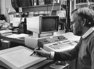
Mike Waterfield at the computer, ca. 1985.
(Photograph courtesy of CRUK London Research Institute Archives.)
The Waterfield laboratory protein sequence database was not the Dayhoff Atlas of Protein Sequences, but a far superior product, NEWAT, first described in 1981 by Russ Doolittle, a protein chemist working in San Diego. Doolittle’s main interest was in molecular evolution, comparing protein structures across different species, and he had gotten so tired of the Dayhoff database’s limitations that he decided to make his own. Because he was in charge of childcare most afternoons after school, he roped in his 11-year-old son Will to do the typing, with his secretary Karen helping with proofreading. Together, the three of them scoured the literature for the sequences of every interesting protein and put together a simple search algorithm so that new proteins could be compared with existing ones. As word spread, Doolittle received hundreds of requests to run protein sequences against NEWAT. In the beginning, he ran the searches himself and then notified the interested parties if there were any matches, but eventually, searching was taking up so much time that he began to give out copies of NEWAT on tape. Not surprisingly, he was a little conflicted about parting with his precious database: “Certainly the first few times, I gave it a lot of thought, because we had gone to a lot of trouble to put this together and now anybody could do what I could do—it was like giving away the golden wand. On the other hand, scientists are very sensitive about their image, and I would never want anyone to think I was the sort who wouldn’t share. I also realised that they weren’t likely to keep it up to date so I’d always have a little advantage over people. So I did it willingly and after a while it just became routine” (BBC 1983).
On 4 May 1983, Mike phoned the Doolittle lab and got through to Karen, Doolittle’s secretary, who, once Doolittle had cleared it, sent a tape of the latest version of NEWAT to London. It was eagerly awaited because the PDGF project had arrived at the point where the protein was pure enough to be entrusted to Nick Totty and Geoff Scrace, the guardians of the sequencers. Two years on from the start of the collaboration, the lab was finally starting to get some very good data.
The tape arrived and was handed over to Peter Stockwell, who loaded up the new database, typed in the PDGF sequence, and set the sequence comparison programme going. What came out was truly remarkable. Human PDGF was an almost perfect match to p28sis, the product of the v-sis oncogene of simian sarcoma virus, and the growth factor and cancer fields had finally, irrevocably fused.
The realisation that p28sis was PDGF was the keystone completing the bridge of data that led to the understanding of how growth factors contributed to cancer. Once it was discovered, it seemed blindingly obvious that, rather than going to the trouble of evolving a new alien growth factor, an RNA tumour virus could just steal a normal cellular growth factor and use it to force cells to proliferate uncontrollably. There was ample precedent for the retroviral theft of cellular genes, but nobody before had figured out the true function of any of the unwilling captives.
Mike recalls vividly what happened: “When Peter found it he came in from the computer and showed me, and we were tremendously excited, because one just doesn’t find this sort of observation unless it is something extremely significant; the degree of identity we found was very unusual. I ran out to talk to a couple of my friends who were virologists, to show them and confirm my feelings about the significance of this observation, and of course they immediately said yes, this was something that was extremely important and interesting.”
Amidst all the excitement, Mike also realised that he and his colleagues would really need to get moving: “The statement ‘p28sis is PDGF’ meant everything to almost every molecular biologist involved in the business. It was like a newsflash, so should we tell everybody, or what should we do? We decided we should write the paper describing our observations and at the same time try and complete the experimental work supporting our observations.”
Getting scooped under such circumstances is the gnawing fear of all scientists, and in the 27th May issue of the journal Science (confusingly published on 20 May), Mike saw with sinking heart an article entitled “Human Platelet-Derived Growth Factor: Amino-Terminal Amino Acid Sequence,” whose authors were Harry Antoniades, one of the Boston PDGF gang, and Mike Hunkapiller, Lee Hood’s right-hand man at Caltech. However, feverish scanning of the paper revealed the unbelievable—Antoniades and Hunkapiller had completely missed the p28sis match. The game was still on, but only just; with both sequences out in the public domain, someone else was bound to find the match in very short order. Mike called Peter Newmark, the biology editor of Nature, Science’s great rival, to ask if he was interested in rapidly publishing a paper that was not only very important but would make Science look a bit silly. Newmark, not surprisingly, said that he was. Mike sat down and wrote a draft of the paper at his dining table in Gospel Oak and then did something that in retrospect was extremely brave, or possibly foolhardy: He and Sally went on holiday to Rhodes, to the little village of Lindos, famed for its ancient acropolis, but not big on telephonic access to the outside world.
At about the time that Mike and Sally were taking their first stroll around Lindos, Russ Doolittle in San Diego was reading Antoniades and Hunkapiller’s paper as some light Saturday morning home entertainment. Being who he was, he too ran a database search with the PDGF sequence and got the same startling result that the Waterfield lab had seen a fortnight earlier:
It was extraordinary. It was a magic moment for me because not only did I find something that matched, but I knew the match. I was familiar enough with this area of biology that the impact was tremendous. It was very interesting, and I’ll never have another moment like that, I’m sure. I took several deep breaths and I had a number of different thoughts. I must confess, among them was “I wonder if anyone else knows this?”—I probably hoped that no one did. And I reflected on who might know. The article had said in it they had conducted a limited search. It wasn’t quite certain what that meant, but I thought “well, if somebody else knows, they know, but in the meantime, I’ll certainly tell people what it is” (BBC 1983).
As was his standard procedure when he found NEWAT matches, Doolittle wrote to Stu Aaronson, whose lab had reported the v-sis sequence, and Hunkapiller and Antoniades. The two groups quickly got in touch, and after some frantic work to extend the PDGF amino-terminal sequence, started preparing a very short paper to be submitted to Science. However, the new paper had acquired several extra authors, one of whom, Lee Hood, was not noted for his ability to keep scientific secrets. Knowing that he was due to give a talk at a big conference in San Francisco on 3 June, he called up Russ Doolittle and asked him if it was OK to mention the PDGF–p28sis homology. Doolittle reacted with commendable mildness, because it was his find, and really, he should have been the one to reveal it to the world:
I wasn’t sure if I minded or not to tell the truth, but I said, “if it’s fine with everybody else.…” I was concerned, I must tell you, as we hadn’t written anything down about this yet, and without having any idea there were any competitors, it just seemed to me indiscreet to be mentioning it before you had published anything or at least had submitted something. At any rate I suspected all the others said the same thing, that it was OK with everybody else, and he mentioned it, to a packed and overflowing crowd I hear (BBC 1983).
Sitting in the second row of the conference hall for Hood’s talk was Tom Deuel, Mike’s collaborator. It must have been an awful moment for him, although his clinician’s ability to dissemble comes to the rescue: “My feelings were very mixed. I was surprised, I was disappointed, I was excited. It was very nice, in the sense of seeing our finding confirmed by others … [but] we were of course disappointed. We wanted to be the first ones to have it presented to the public” (BBC 1983).
Once out of the session, Tom phoned the Waterfield lab and discovered to his horror that Mike was off on holiday. Once he’d told the lab what had happened, they dispatched a telegram to Mike, simply stating “PLEASE CALL LAB.” From amongst the feta cheese and olives in the local supermarket in Lindos, Mike did just that, and told Paul Stroobant to get the paper off to Peter Newmark at Nature as quickly as possible. The next day, another telegram arrived, this one simply announcing “NEWMARK READY PROBABLY MONDAY PM. FOUR WEEKS OUT.”
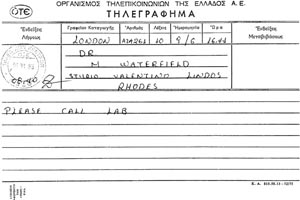
Holiday disruption: The telegram Mike received from his laboratory.
(Courtesy of Mike Waterfield.)
The American paper, with Russ Doolittle as first author and a single figure showing the match between PDGF and p28sis, was submitted to Science on 3 June, and accepted on 12 June. Mike’s lab, still in his absence, got their paper, a rather longer affair, to Nature on 14 June, where it was accepted three days later. The speed of both journals was completely unprecedented, and the papers were eventually published by Science on 15 July (although, as mentioned above, this really meant 8 July), and by Nature on 7 July, a testament to how intense the competition between the two journals was at the time. However, by the time of the papers’ publications, both camps were enveloped in a media storm, thanks to an indiscreet and overeager journalist who had heard Hood’s talk and written it up for the 18 June issue of a magazine, Science News.
Shortly after Science News broke the story, Russ Doolittle received a phone call: “A reporter from the Los Angeles Times called up here, and said ‘See here, I see your name mentioned in this thing. Would you like to tell me about your role in it?’ Well … our public relations people here said ‘Fine, talk to reporters. It’s too late now—there’s no embargo any longer.’ ” Doolittle’s press contact also told him about the rival paper:
He said, “By the way, I just talked to someone at the Salk Institute and they told me there are some people in England that have discovered the same thing.” And he told me the name of the person, which I confess I didn’t recognise at the time. And, well, I guess … there was some disappointment to find that somebody else was on top of it so quickly. On the other hand, it happens, and that’s the way it is.… It wasn’t until later that evening that it dawned on me that the name that had been told to me was familiar, and I went through my records and found that Dr Waterfield had requested one of the tapes. It was ironic … I guess I was relieved that our database had played some role in their discovery also, and that sort of made it nice again I guess.
Would he have liked Mike to have informed him of the homology? “I’ve seen it both ways, and yes, I would like to have known, but I also quite understand why he didn’t tell anybody.… Given the nature of the work they were doing, it seems to me that most people would have behaved the same way. The one thing I think I wouldn’t have done if I were he, is that I would never have gone off on holiday until I had finished the task!” (BBC 1983).
Once the story was out in the Los Angeles Times, it was picked up by the rest of the U.S. media, and there was no holding it. Mike, finally back from his holidays, got a call: “We were asked by the BBC World Service if we had done this particular piece of work because they had seen it in the Herald Tribune.… I think we would much much rather have held a well-ordered press conference at which we could fully explain everything, but we did the best we could.”
The ICRF, with a nonexistent press office and limited experience in dealing with the media, put out a press release that Peter Newmark subsequently described as “not cautious. It did not mention the fact that the only strong link of the discovery to cancer was in the case of tumours of monkeys caused by simian sarcoma virus. It claimed categorically that PDGF ‘produced by the oncogene in excessive amounts … leads to uncontrolled growth in … bones and other connective tissue’ and that drugs can now be devised to block the effects of PDGF and ‘perhaps stop cancerous growth’” (Newmark 1983).
Mike, without the protection of a public relations department, found himself besieged by a press pack that had scented a miraculous cure for cancer. At work, it wasn’t too bad: “It got picked up when there wasn’t a lot of news. ICRF was invaded by a lot of newspapers including some journalists from Brazil who were there to cover a football competition. We had these guys come in the lab and they were filmed. We dressed them up in lab coats but they were football correspondents!” At home, however, it was a lot more alarming. Mike and Sally were doorstepped aggressively by reporters: “It was quite upsetting because I was very naïve and open about it and then got burnt—Sal and I ended up with our picture on the front of the News of the World which was taken because the guy was banging on our door in Oak Village, and we let him take the picture in order to make him go away.”
Although many of the papers managed to produce a reasonably accurate account of the work following the press release, others did not. Mike and Sally’s nemesis, The News of the World, was especially irresponsible, leading with the headline: “Cancer Jab on the Way,” dubbing poor Mike a “superboffin” and quoting him as saying that “within the year, or at least by this time next year, we will find a drug to halt some kinds of cancer. It would be nice to say a quick pill will solve the problems, but I don’t think that’s so.… I think it will have to be an injection.” The story ended with the tale of a little girl, Victoria Hart, who had just died of bone cancer, and implied that her cancer might have been cured by the mythical cancer jab. It was a monument to the reporting style that led to the paper’s eventual closure in 2011.
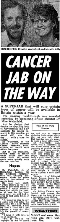
Article from News of the World, 3 July 1983, © NI Syndication.
Mike emerged bruised and blinking from his ordeal by media, and life returned to some semblance of normality by the autumn of 1983. Work on PDGF continued, but other things were also quietly afoot in the Waterfield lab. In line with his long-standing interest in membrane proteins, acquired in his days at Caltech working on immunoglobulins, Mike had been running another project in parallel with the PDGF work: to purify and sequence the receptor for Epidermal Growth Factor, the opener of baby mice’s eyes.
Nearly 20 years on from Stan Cohen’s original isolation of EGF, addition of the protein to cells was known to induce the export and import of many substances, stimulate cellular metabolism and proliferation, cause changes in cell shape and behaviour, and activate DNA, RNA, and protein synthesis. How EGF did any of these things was more or less a mystery, but binding of EGF to its cellular receptor was an absolute requirement. The receptor was an intriguing molecule. It had a tripartite structure, with an extracellular EGF-binding domain, a transmembrane region, and an internal domain, which the Cohen lab showed in 1980 possessed tyrosine kinase activity, the ability to identify the amino acid tyrosine in a protein and stick phosphate groups on it. The receptor’s tyrosine kinase activity was tightly regulated—EGF binding was the “on” switch, whereas in the absence of EGF, activity was firmly off.
The finding that the EGF receptor had tyrosine kinase activity was the first biochemical link between oncogenes and growth factors, giving substance to the theories suggesting that both might work to stimulate cell growth through similar pathways. Protein kinase work had become obligatory in the oncogene world after the laboratories of Mike Bishop, Harold Varmus, and Ray Erikson discovered in 1978 that v-Src, the oncoprotein of Rous sarcoma virus, had protein kinase activity. Phosphorylation of proteins can alter their functionality dramatically, and the idea that oncogenes might be able to tamper with such a fundamental control mechanism was immensely attractive. Accordingly, everyone looked to see if their pet protein could join the kinase club. It was on such a quest that tyrosine kinases were discovered by Tony Hunter in San Diego, during the three-way race to define the function of the middle T oncogene of polyomavirus. Hunter and Bart Sefton went on to show that v-Src and its cellular progenitor c-Src were both tyrosine kinases, and using their assay, tyrosine kinases started popping out of the woodwork in all directions, including in the growth factor world. The EGF receptor was the first so-called Receptor Tyrosine Kinase to be categorised, but was soon joined in 1982 by the receptors for insulin and PDGF, suggesting the existence of a common pathway for intracellular signaling by some growth factors. The Waterfield lab’s discovery that PDGF was the p28sis oncoprotein was the punchline for the whole story to date, showing how this normal pathway could be subverted.
Although the EGF receptor had an extremely trendy function ascribed to it, it had yet to be sequenced, and taking on such a challenge was exactly what Mike and his lab loved. Working in their favour was the fact that they could tap an unexpectedly rich source of protein; A431 cells, a human squamous carcinoma line, had been shown by Joe DeLarco to carry millions of EGF receptor molecules, as opposed to the normal number of a few thousand. This had not been overlooked by the rest of the field, and the Cohen lab had begun purifying the EGF receptor in the late 1970s by running A431 membrane extracts down an affinity column to which EGF protein had previously been chemically coupled. Under the appropriate conditions, the EGF proteins could hook their receptors out of the extracts as they passed through the column, whilst everything else simply flowed past. The EGF receptor molecules could then be washed off the column by changing the binding conditions. However, even the great Stan Cohen had been unable to get any sequence by this method because the yield was too poor. At the ICRF, Mike, looking around for a better alternative, got talking to his next-door neighbour, Peter Goodfellow.
Peter Goodfellow’s main interest was in human genetics, as will be related in Chapter 8, but in the early 1980s, freshly arrived at the ICRF, he was much in demand for another of his skills—making monoclonal antibodies. As shown to great effect for SV40 large T and p53, monoclonal antibodies were and are by far the best tools for pulling specific proteins out of complex mixes, and after having a long discussion with Mike, Peter came up with a very cute way of making a monoclonal antibody against the EGF receptor. Rather than dithering about making huge quantities of purified receptor and injecting it into mice in order to get an immune response, why not simply immunise mice with A431 cells? There was a very good chance that that would work just as well, given that the cells were fairly bristling with receptors, and it would be an awful lot easier and faster. Monoclonal antibody–producing cells (hybridomas) could then be made, and the antibodies they secreted tested for their ability to block EGF binding to A431 cells; if the monoclonal was specific for the EGF receptor, it would sit on it, thereby hiding it from EGF and giving a negative result in an EGF-binding assay. The trick worked rather well, and a joint paper describing EGFR1, the first specific anti-EGF receptor monoclonal antibody, was published in September 1982, with the Waterfield lab, in particular Mike’s postdoc Brad Ozanne, doing the binding assays, and George Banting from Peter’s lab covering the antibody work.
In October 1982, Mike acquired a new graduate student, hired to join the EGF receptor sequencing project. Julian Downward arrived at the ICRF trailing a veritable wagonload of British establishment baggage; he’d been to Eton and Cambridge, and his Dad was a Major General in the British Army. Fortunately, none of these things managed to obscure the fact that Julian was both extremely nice and so self-effacing that it was easy not to notice just how good a scientist he was. He’d come to the ICRF on the recommendation of his lecturers, who’d characterised the place as a land of milk and honey compared with their impecunious laboratories in the windswept wastes of the Fens, but when he arrived Julian found that things weren’t much different to the Cambridge Biochemistry department:
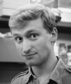
Julian Downward, 1984.
(Photograph courtesy of Peter Parker.)
I thought that everything would be shiny and people would be walking around in designer outfits or something, but it was actually fairly similar to Cambridge. In Mike’s lab, I remember that a lot of people had beards, so it had a sort of chemistry feel to it. He had these two senior technicians, Geoff Scrace and Nick Totty, who were very beardy, and their job was to run all these sequencers. It did seem quite impressive—the machinery was all very impressive. I got a tremendous sense of enthusiasm from Mike, which was very endearing. He said cancer was bound to be a subversion of the way normal growth mechanisms worked and this was the way to find out.
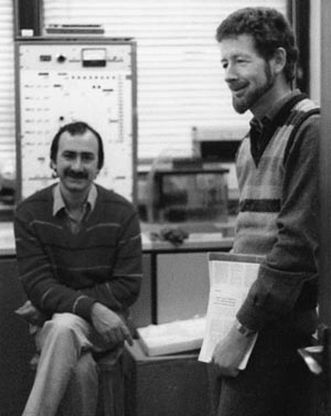
“Very beardy.” Nick Totty and Geoff Scrace, ca. 1984.
(Photograph courtesy of Peter Parker.)
Mike realised quite fast that he’d lucked out with his new student:
He came in on day one and decided on the project and said, “Right, I want to get started.” I said, “What help do you need?” and he said, “None, I’m just going to get on with it.” And he quietly worked his butt off! I think [he was] someone who was just focussed, fascinated and extraordinarily capable—he obviously had a CV that said he’d done rather well at Eton and rather well at Cambridge—I think he’d passed out first in both places. Remarkable guy, and he was also quite a funny mixture. Now, I don’t regard him as being socially wild … he’s cool and calm. But in those days during his PhD he was really wild at times! We used to go down to conferences at Wye College, and I had some amazing times with Julian. Over the years he’s settled down a bit—maybe too quickly!
The EGF receptor project had been going for a little while, with a postdoc, Elaine Mayes, doing protein stuff using A431 cells, and a PhD student, Nigel Whittle, trying to clone the EGF receptor gene directly. Julian’s job was to work with Elaine on receptor purification, but instead of using A431 cells, Mike wanted him to try a different source. Some years back, it had been noticed that there were lots of EGF receptors on placental membranes, and Sally knew a gynaecologist at Queen Charlotte’s Hospital. Putting these two facts together led to the 22-year-old Julian being sentenced to collect fresh afterbirths from the new mothers of Hammersmith, an experience he remembers vividly after more than quarter of a century: “Getting it from placenta was just a complete nightmare. I used to go over and hang around all day waiting for these things to come. I’d sometimes do a few at a go depending on what was available. I would be waiting in the sluice room with loads of ice. It wasn’t very nice at all!”
Once the placenta had been transported back on the tube, Julian would settle in for a long evening in the lab:
Placenta’s a bit looser than liver but it’s that sort of thing. The first step in the process was you’d cut off the gross membranes and then with all the meaty bits you’d cut them into little chunks in cold PBS [phosphate-buffered saline] and put them into a blender. Then you’d spin that stuff down and put it into a low osmotic strength buffer for an hour. The idea was that these villi would bleb off and so you’d get this preparation of villi, and those were very rich in EGF receptor. So you would end up with a nice clean membrane fraction from the surface of the syncytiotrophoblast. Once you got those membrane preps you could then store them and freeze them. Usually on those days if I could get away from the hospital about four in the afternoon I would be finished by midnight, but if it got much later than that it would just go on and on. And there was loads of mercaptoethanol and everything else and it stank.
After a few months of dicing and slicing, Julian was getting small amounts of pure placental EGF receptor and had also discovered that Peter Goodfellow’s EGFR1 monoclonal antibody was a far better purification reagent than were the old Cohen-style EGF-binding columns. However, in the absence of a West London population explosion, Julian had also realised that it was going to be very hard to get enough placenta to make sufficient EGF receptor to sequence. The A431 cells were clearly the way to go.
Putting placenta on the back burner was surprisingly easy, thanks to the PDGF and p28sis story going nuclear about six months after Julian’s arrival. Many of the people in the lab, including Elaine and Nigel, who were both tired of fighting a losing battle with the EGF receptor, dumped their projects in favour of PDGF-ology, and Julian was left to carry on by himself: “I did think about jumping too, but then I thought ‘Well, actually this is quite an interesting project in its own right, and it would make life a lot simpler if it was just open to me.’ So I started doing a lot more on the A431 cells. I started producing huge amounts of cells with the cell culture people here—I started getting them to do eighty 2.5-litre roller bottles a week. The cells grew like weeds and you got vast amounts of cells and they had 2 million receptors per cell—the amount you got was just unbelievable. Suddenly it all started to come together pretty well.”
Despite Julian’s having to deal with a bathtub’s worth of cells every week by himself, getting lots of EGF receptor was in the end quite straightforward, thanks to the EGFR1 magic monoclonal antibody. He even managed to get something from the placental preps, which he kept doing, “because I just wanted to feel that the placenta had led somewhere.” The only real problem was the cold room, where Julian spent a large part of his time mollycoddling his protein. Keeping warm whilst doing experiments in there meant having to dress as though it were snowing, even in high summer, but as an additional hazard, the fourth floor cold room was also liberally anointed with fresh cow brains. Peter Parker, a recently arrived postdoc with Mike, was trying to purify an enzyme called protein kinase C from the brains, so fellow users had to breathe a pungent aerosol of brain and mercaptoethanol, singeing their sense of smell and potentially exposing themselves to mad cow disease all in one go.
By the end of the summer of 1983, as Brazilian football reporters, assorted gutter journalists, and BBC film crews surged through the laboratories in pursuit of his superboffin boss, Julian had got enough protein together to put onto the sequencer, under the watchful eye of Nick Totty. He had also acquired a temporary collaborator from the Weizmann Institute in Israel, a PhD student called Yossi Yarden. Mike had set up a collaboration with Yossi Schlessinger, Yossi Yarden’s boss, thinking that he might need another source of EGF receptor should Julian not get much from A431 cells. The Israeli source was superfluous to requirements by the time Yossi arrived, but the few weeks of additional company weren’t unpleasant:
Yossi is very taciturn and he’s got this big hearing problem from when he got blown up while he was in the Israeli paratrooper brigade doing his national service. He was a nice guy, very straightforward. When they finally got their stuff onto the sequencer it didn’t really give anything that was obviously the EGF receptor. What we did get out of it was ubiquitin, which at the time we just thought was some contaminant. Actually, it probably was interesting, as the receptor is ubiquitinated, and now there’s a whole industry about ubiquitination of the EGF receptor.
Yossi eventually returned to Israel without getting anything sensible in the way of sequence, but went down in lab mythology for his ability to fall asleep instantly and anywhere, as Mike remembers: “He used to occasionally spend the night sleeping on the bench in the lab because when he’d been in the Israeli army he used to run miles and miles with a pack and rifle across the desert—he always said he could sleep even when he was running. He was a wonderful guy—quiet, determined.”
By the winter of 1983, Julian had around 250 amino acids-worth of sequence from the EGF receptor, but the protein had had to be chopped into smaller peptides for sequencing, and there was no way of knowing how the resulting short sequences fitted together; the words were all there, but the story they told could not be deduced. This wouldn’t matter if the peptide sequences matched something in the Doolittle database, but disappointingly, when Peter Stockwell ran a search, there were no homologies to anything. It looked as though Julian would be in for the long haul of figuring out what possible DNA sequences coded for the peptides and using these to go on a fishing expedition for the EGF receptor gene in a human DNA library. The methodology for all this was still very new, and so Mike roped in a friend of his to help. Axel Ullrich from Genentech, the company that Herb Boyer and Bob Swanson had set up to exploit recombinant DNA technology, already worked on EGF, was a cloning expert, and was very keen to get on board. However, before Ullrich could get started on the project, Julian hit pay dirt:
A paper had come out from a guy in Japan [Tadashi Yamamoto and colleagues] who had been sequencing the v-erb-B oncogene from AEV [avian erythroblastosis virus]. So Geoff Scrace put the sequence into the database. Every time Geoff updated the database I’d just do a sequence search without relying on Peter Stockwell. I used to do it late at night because that’s when Peter went home and I could get onto the computer terminal (there was only one computer terminal!). If I did it when he was around he was really grumpy and he used to shout at me because I was disturbing what he was doing. So I would wait till he went home.
I just did it one evening—every couple of weeks I’d check—and then suddenly saw these things all lined up. You know, the first one, I thought maybe that’s chance, but just more and more kept coming up on the screen. There were about fifteen to twenty sequences and about half of them were lining up. They all just started popping out and it was very obviously a big deal—the matches were perfect—I mean not totally perfect but every peptide aligned—90% or so homologous. The v-erb-B oncogene is truncated—it’s lost most of the amino terminal part but it’s got the transmembrane region and most of the intracellular bit. So it was about half of the EGF receptor represented there.
So then I called Mike at home—it was probably about nine in the evening in early December. Mike realised very rapidly this was something important so he came in with Sally and we spent the next few hours tinkering around with these sequences, and then he drove me home.
Discovering something quite so important after only 15 months as a PhD student is something most scientists can only dream of, and Julian was well aware of the significance: “At the slow times in the work before that I had thought how difficult it was to achieve anything—I remember sitting up in the library reading copies of Nature and thinking ‘there’s so much work here—how can anybody achieve all this in a PhD?’ But then when it all happened I knew it was a big deal. And then Paul Stroobant said ‘This is something you should savour because probably you’ll never do anything as important as that again.’ It was pretty reasonable advice. I appreciated how important it was.”
Julian and Mike spent Christmas 1983 writing up the work, and submitted a paper entitled “Close Similarity of Epidermal Growth Factor Receptor and v-erb-B Oncogene Protein Sequences” to Peter Newmark at Nature in mid-January 1984. Perhaps fearing a repeat of the PDGF saga, Newmark accepted the paper in five days and published it a fortnight later, but this time, there were no nasty scientific surprises in store.
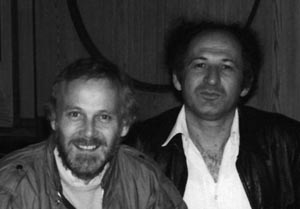
Mike Waterfield and Yossi Schlessinger, ca. 1985.
(Photograph courtesy of Martin McMahon.)
Having been thoroughly shaken by the attentions of the press the previous year, Mike made sure that he wasn’t in London when the paper came out: “I waltzed off to Israel and gave two big lectures at Tel Aviv university, and had a great time!” Sally was left behind to fend off the journalists by herself, because Julian was also absent. Mike had delegated a rather important task to him:
Mike got invited to a UCLA meeting in Steamboat Springs [a ski resort in Colorado] called something like “Genes and Cancer,” and for some reason he didn’t want to go. So he said (looking back on it, it’s fairly unbelievable!), “I’ve got a 23 year old graduate student, he’ll be perfect to give a talk at this major meeting.” It was the first talk ever that I’d given. We put together a presentation and I actually got a lot of advice from Mike Fried. Mike decided that Mike Waterfield was not taking this sufficiently seriously and I would be eaten by the lions unless I was highly trained up to give this talk. He was very helpful and gave a lot of advice going over this talk and how to present it.
So I headed off to this meeting, which was shortly before the paper came out in the middle of February, and gave this talk, which made a big splash. Tony Hunter was an organiser of the meeting and I remember he invited me to dinner with him and some other people in this fancy restaurant in the resort and I thought this was amazing because I knew Tony Hunter’s work from my undergraduate days, and subsequently.
Tony Hunter, the kinase king, remembers Julian’s talk at the Steamboat meeting very well. He’d heard on a visit to London in December 1983 that the Waterfield lab had a lot of EGF receptor sequence but the homology with the v-erb-B protein had not yet been noticed. When Julian got up to speak, his training in the Mike Fried Seminar Bootcamp paid off, and he knocked the collective socks off his audience, including Tony: “I recollect that he was very British and proper, and amazingly poised and confident for a graduate student—he held his own at the UCLA meeting, and at a meeting in Kyoto he came to instead of Mike in November 1985.” Julian doesn’t recall being particularly worried by the attention: “It was just exciting, really. I didn’t feel out of my depth—I feel much more out of my depth now than I did then. At the time I felt I knew more about this than anyone else in the world and I think that was probably true.”
In May 1984, the Waterfield lab had another Nature paper to celebrate, this time with a crowd of collaborators from the Ullrich and Schlessinger labs, in which the complete coding sequence of the human EGF receptor gene was presented. Comparison of the sequence of the cellular gene with its oncogenic relative v-erb-B showed that the oncogene was truncated, deleted, and mutated in such a way that the protein it encoded was no longer regulated by EGF binding, but instead was permanently switched on, pushing cells to grow uncontrollably. This, together with the PDGF story, was the clinching evidence for a truth that is now ingrained in the collective consciousness of cancer biologists: oncogenes cause tumours because they encode mutationally activated components of normal cellular growth pathways. As befits their status, the three papers that the Waterfield lab published in that one remarkable year have been cumulatively cited around 6000 times. The perfect combination of state-of-the-art database mining, protein purification, and sequencing, matched with Mike’s outstanding eye for a good problem and a good student, had paid off in spades.
In cancer, there are now many examples of mutated or overactive growth factors and receptors causing particular human tumours, and numerous drugs have been developed that target such molecules, many based on neutralising monoclonal antibodies such as Peter Goodfellow’s EGFR1. Mutations in the EGF receptor itself have been found in multiple cancer types, and several effective EGF receptor–inhibiting drugs are now in use in the clinic. One of the best known of these is the therapeutic antibody Herceptin (trastuzumab), a standard and effective part of first-line breast cancer treatment developed at Genentech by, amongst others, Axel Ullrich.
Thanks in part to the Waterfield lab and their collaborators, signal transduction (how extracellular messages affect intracellular events) became the fastest growing field in biology in the 1980s. However, although it was evident that deregulation of growth factors and their receptors could cause cancer, the pathways leading from receptor activation into the cell petered out in a dense thicket of uncertainty. One obvious line of inquiry, trying to work out what proteins the receptors were able to phosphorylate, proved to be annoyingly circular, because their only convincing substrates were themselves. Educated guesswork, seeing whether other known oncogenes could be activated by growth factors, was deployed with more success. In 1984, a paper by Tohru Kamata and Jim Feramisco gave the first vague hints that addition of EGF to cells somehow stimulated the activity of the Ras oncoprotein. This tenuous link strengthened into reality in 1986, when Dennis Stacey and colleagues used antibodies blocking Ras activity to prevent transformation by receptor tyrosine kinases. Ras clearly had something to do with receptor signaling, but whether it lay directly downstream from the receptors or was on a parallel pathway was still an open question.
Ras had a great deal of previous form as an oncoprotein. In 1979, Bob Weinberg’s lab at the Whitehead Institute in Boston showed for the first time that there were human genes that could cause cancer when mutated. Using the new technique of DNA transfection, whereby foreign DNA could be introduced directly into cells, they showed that a fragment of DNA from a human tumour was able to transform mouse NIH3T3 tissue culture cells. Other groups began to see the same results using DNA from other tumour types, and together, they realised that the fragments of human DNA they were isolating all contained the H-ras gene, the cellular homologue of v-H-ras, one of two closely related retroviral oncogenes from rat sarcoma viruses. Ras family proteins are attached to the inside of the cell membrane and belong to a larger protein family, the GTP-binding proteins. Such proteins are able to bind guanine triphosphate (GTP), a product of cellular metabolism, and convert it to guanine diphosphate (GDP). It was known that the mutations that converted normal Ras proteins into oncogenes caused them to stick in the GTP-bound activated state so that they were switched on the whole time, but here the trail ran out. Ras sat in solitary splendour, with its upstream and downstream partners still shrouded in mystery.
In 1987, after writing up probably the most spectacular PhD ever done at the ICRF, Julian Downward was sucked into the black hole surrounding the Ras proteins when he started a postdoc in Bob Weinberg’s lab. It was fortunate that he’d had such a good experience as a PhD student, because his postdoctoral work was nowhere near as successful, consisting of mostly fruitless attempts to work out what the trigger for activation of normal Ras proteins might be: “I spent most of 1987-9 throwing EGF and similar growth factors at cells and looking at the GTP bound to Ras, but never saw any effects, and many others had the same experience. It was a technical problem and in fact was not completely solved for a long time … the stimulatory effects were just too small in most of the cell systems to rise above the noise inherent in the assay.”
In 1989, Julian was recruited back to the ICRF to run a laboratory in his own right, and arrived still shackled to his annoyingly intractable postdoc project. Although Mike had moved on to become Director of the Middlesex Hospital branch of the Ludwig Institute for Cancer Research, Julian still found a sympathetic audience for his Ras-related problems: his fellow group leader Doreen Cantrell, who was (and still is) a leading T-cell immunologist. T cells are a vital part of the immune system, binding to foreign antigenic invaders and activating the body’s powerful defences against disease. As an experimental tool for studying signal transduction, they are also extremely attractive because they can be easily purified and fooled in vitro into activating themselves if they are fed mock antigens. Doreen suggested to Julian that her T-cell system would be an ideal place to look at what was activating Ras. The shift in perspective, as can often happen in scientific research, broke the logjam. In just four months, the collaboration succeeded in doing what Julian had been labouring over in Boston. Working together, the two labs showed for the first time what everyone had suspected but had been unable to prove: Ras could be activated rapidly and strongly in response to extracellular signals stimulating a receptor, in this case, the T-cell receptor, the cell surface molecule on T cells to which antigen binds.
Once Ras activation in T cells had been shown, it could be used to tune up the problematic GTP-binding assay to detect what else switched Ras on. The signal-to-noise ratio was improved to the point that many other receptors were shown to turn Ras on, including Julian’s old friend the EGF receptor. The race was now on to join up the dots between the receptors and Ras. Julian and his postdoc Laszlo Buday realised that work performed genetically dissecting Ras signaling in fruit flies could be put together with biochemical clues from mammalian cells to produce two likely candidates for the missing links, the Sos and Grb2 proteins. Sos (Son Of Sevenless; the Drosophila field is inordinately fond of giving genes daft names) was known to act downstream from the fly EGF receptor gene and upstream of fly Ras, and there was also good reason to think that it was a guanine exchange factor, able to catalyse rapid exchange of GDP for GTP, and therefore a prime candidate for activating Ras. Grb2 had been proposed to be an adaptor protein, acting as a link between the EGF receptor and intracellular signalling components, and there was indirect evidence implicating it in Ras activation. Leaving T cells for fibroblasts, Julian and Laszlo wallowed enthusiastically in radioactivity for a few months, labelling cells and using antibodies to try to immunoprecipitate Ras protein along with Grb2 and Sos, and then doing in vitro biochemistry to show that the interactions they detected resulted in Ras becoming activated. Their efforts successfully showed that the EGF receptor, when activated by EGF binding, recruited Grb2 to its intracellular tail, that Grb2, in turn, bound Sos, that Sos had hold of Ras, and that all together, this enabled Ras to be activated. This work, together with a similar paper from ICRF alumnus Tony Pawson’s lab in Toronto, defined the missing upstream links.
But what lay downstream from Ras, between it and the genes controlling the cell cycle? Part of the answer lay with another proto-oncogene, c-Raf-1. Raf1 protein is a kinase, and there was indirect evidence linking it with activation of Ras, because disruption of Raf1 activity had been shown to block Ras action. The Downward lab used a piece of Raf1 as a biochemical bait to see whether it was able pull down Ras protein and showed that, indeed, Ras and Raf1 interacted, but only when Ras was in its activated, GTP-bound state. This was a crucial interaction to map, because downstream from Raf1 lay the mitogen-activated protein kinase (MAPK) cascade. Kinases belonging to the MAPK family are used throughout evolution to control the cellular responses to external signals such as growth factors, nutrient status, and stress or inductive signals, and MAPK had just been shown by Richard Treisman’s lab, just upstairs from Julian at the ICRF, to regulate transcription factors. The extracellular signalling pathway had reached the nucleus, and ICRF scientists had played a big part in illuminating many of its twists and turns.
There is one further chapter to the story, as became clear to Julian that not all of the things that Ras was known to be able to do could be explained by it simply activating the MAPK pathway. Another protein, phosphatidylinositol-3-kinase (PI3K), had been shown in 1992 to interact with Ras in vitro, and PI3K was known to be activated in response to the same stimuli as Ras. Julian’s lab, in collaboration with Mike Waterfield’s at the Ludwig Institute, showed in 1994 that activated, GTP-bound Ras bound directly to PI3K, and that this interaction was likely to result in PI3K being activated in turn. The work placed Ras at the apex of two downstream signalling cascades, both of which are vitally important for cell growth.
The basic mechanism of how extracellular signalling works has been fleshed out in the intervening years such that we now have a very detailed map of signal transduction in both normal and cancerous cells. Growth factors and other extracellular stimuli sit at the top of a host of pathways governing cell growth and differentiation, and the molecular wiring diagrams for different cell types are varied and complex. Scientists are turning towards systems biology to model how perturbations in one part of a cell’s wiring change the outcome for the cell, and signal transduction research is becoming rigorously quantitative. Fresh insights into the processes involved are won at huge cost; there is no low-hanging fruit left to pick in the garden of molecular biology. The excitement of the early days, when huge leaps in understanding were almost commonplace, has gone, to be replaced by the tramping feet of an army of scientific foot soldiers marching towards the ultimate goal—complete knowledge of a cell and the medical exploitation of that knowledge. They will get there, and there will doubtless be other kinds of fun along the way, but there will never be another time when one lab equipped with a small computer, a fancy sequencing machine, and some great ideas could change our view of the molecular landscape so profoundly.
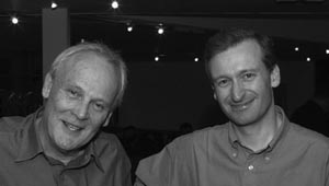
Mike Waterfield and Julian Downward, 2002.
(Photograph courtesy of CRUK London Research Institute Archives.)
Further Reading
Cohen S. 2008. Origins of growth factors: NGF and EGF. J Biol Chem 283: 33793–33797.
A very clear account of Stan Cohen’s early life and work.
Levi-Montalcini R. 1988. In praise of imperfection. Basic Books, New York.
Rita Levi-Montalcini’s autobiography. A remarkable book about a remarkable woman
Web Resources
http://blip.tv/portland-press-ltd/mike-waterfield-ludwig-institute-for-cancer-research-ucl-london-2691775 Mike Waterfield in action.
www.asbmb.org.au/magazine/2002-August_Issue33-2/Pehr%20Edman%20Feature.pdf Contains an extremely frank account of Pehr Edman’s personality by his ex-research assistant, as well as an assessment of his life and work.
www.youtube.com/watch?v=wiu7DMjG6RE Russ Doolittle is an enchanting speaker. Watch him lecture.
Quotation Sources
BBC TV Horizon series. Cancer, the pattern in the genes. Broadcast 5 December 1983.
Ibid.
Ibid.
Levi-Montalcini R. 1988. In praise of imperfection: My life and work. Basic Books, New York.
Newmark P. 1983. Oncogene discovery. Priority by press release. Nature 304: 108.
William Dreyer interview by Shirley K. Cohen, 18 February–2 March 1999. Oral History Project, California Institute of Technology Archives, reprinted with permission. http://www.archives.caltech.edu/search_catalog.cfm?results_file=Detail_View&recsPerPage=1&firstRecToShow=72&search_field=&entry_type=NonPhoto&photo_id=&cat_series=oral%20history.